
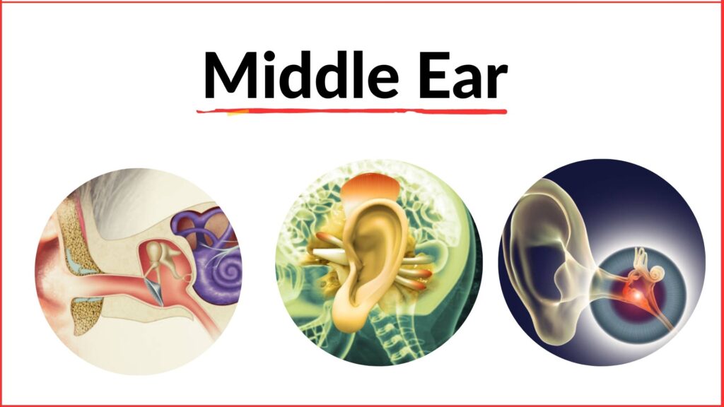
Middle ear : Anatomy, features & function
Middle ear
Anatomy of middle ear
Content of middle ear cavity
Middle wall of middle ear cavity
Functions of middle ear
Describe inner ear
Development of middle ear : Tympanic cavity and eustachian tube from the endoderm of tubo-tympanic recess between the 1″ and 2nd branchial arch.
Anatomy of middle ear :
- The middle ear is also called the tympanic cavity, tympanum.
- The middle ear is a narrow air filled space situated in the petrous part of the temporal bone between the external ear and the internal.
- The middle ear is shaped like a cube.
- Its lateral and medial walls are large, but the other walls are narrow, because the cube is compressed from side to side. Its vertical and anteroposterior diameters are both about 15 mm.
- When seen in coronal section the cavity of the middle ear is biconcave, as the medial and lateral walls are closest to each other in the centre.
- The distances separating them are 6mm near the roof, 2 mm in the center, and 4 mm near the floor.
- Middle ear can be likened to a six-sided box with a roof, a floor, medial, lateral, anterior and posterior walls.
Parts:
- Mesotympanum : lying opposite the pars tensa.
- Epitympanum or the attic : Lying above the pars tensa but medial to Shrapnell’s membrane and the bony lateral attic wall.
- Hypotympanum Lying below the level of pars tensa.
The portion of middle ear around the tympanic orifice of the eustachian tube is sometimes called protympanum.
Communications :
- The middle ear communicates anteriorly with the nasopharynx through the auditory tube, and posteriorly with the mastoid antrum and mastoid air cells through the aditus to the mastoid antrum.
- The middle ear is likened to a sump pit or trap in the sloping course of the aditus to the epitympanic recess and the auditory tube.
Boundaries of middle ear cavity :
- The Reefer Tegmental wall : Thin plate of bone (Tegmen tympani) which separates the cavity from the middle cranial fossa.
- The Fleet or Jugular wall : Thin plate of bone which separates the middle ear cavity from the jugular bulb of internal jugular vein.
- The Anterior or Carotid wall : The anterior wall has a thin plate of bone, which separates the cavity from internal carotid artery.
- The Lateral or Membranes walk : Tympanic membrane and squamous part of temporal bone.
- The Medial or Labyrinthine wall: Formed by thin plate of bone, this separates middle.
- The Posterior er Mastoid wall :
- Descending part of facial nerve.
- Aditus antrum.
- Pyramid.
Medial wall of middle ear cavity :
Features of medial wall :
- The promontory is a rounded bulging produced by the first turn of the cochlea. It is grooved by the tympanic plexus.
- The fenstra vestibull is an oval opening posterosuperior to the promontory. It leads into the vestibule of the internal and is closed by the footplate of the stapes.
- The prominence of the facial canal runs backwards just above the fenestra vestibuli, to reach the lower margin of the aditus. The canal then descends behind the posterior wall to end at the stylomastoid foramen.
- The fenstra cochleae is a round opening at the bottom of a depression posterior-inferior to the promontory. It opens in the scala tympani of the cochlea, and is closed by the secondary tympanic membrane.
- The sinus tympani is a depression behind the promontory, opposite the ampulla of the posterice semicircular canal.
- The process cochleariformis (The bony septum between the canals for the tensor tympani and for the auditory tube is continued posteriorly on the medial wall as a curved lamina called the processs cochlearifornnis).
- Prominence of lateral semicircular canal above the facial canal.
Middle ear cleft :
Definition : Middle ear cleft is composed of the tympanic cavity with its contents, mastoid air-cell system.
Contents of middle ear cleft :
1) Middle ear cavity : Contents of middle ear cavity are-
- Auditory ossicles : Malleus Incus & Stapes.
- Ligaments of ear ossicles.
- Muscles: Tensor tympani & stapedius.
- Vessels supplying and draining the middle ear cavity.
- Nerves: Tympanic plexus, Chorda tympani.
- Air.
2) Eustachian tube
3) Aditus to the antrum
4) Mastoid
5) Mastoid air cell.
Muscles of the middle car :
- Tensor tympani
- Stapedias.
Nerve supply of the middle ear :
- Tympanic plexus
- Chanda tympani
Structures present in the anterior wall of the middle ear cavity :
- The uppermost part of the anterior wall bears the opening of the canal for the tensor tympani.
- The middle part has the opening of the auditory tube.
- The inferior part of the wall is formed by a thin plate of bone which forms the posterior wall of the carotid canal.
- The plate separates the middle ear from the internal carotid artery. This plate of bone is perforated by the superior and inferior sympathetic caroticotympanic nerves and the tympanic branch of the internal carotid artery.
- The bony septum between the canals for the tensor tympani and for the auditory tube is continued posteriorly on the medial wall as a curved lamina called the processus cochleariformis.
- Its posterior end forms a pulley around which the tendon of the tensor tympani turns laterally to reach the upper part of the handle of the malleus.
Function of middle ear :
- It transmits sound waves from the external ear to the internal ear through the chain of car ossicles, and thus transforms the airborne vibrations from the tympanic membrane to liquid bome vibrations in the internal ear.
- The intensity of the sound waves is increased ten times by the ossicles. It may be noted that the frequency of sound does not change.
Internal ear :
Internal ear :
- The internal ear, or labyrinth, lies in the petrous part of the temporal bone.
- Two parts: Bony labyrinth & Membranous labyrinth.
- The membranous labyrinth is filled with a fluid called endolymph.
- It is separated from the bony labyrinth by another fluid called the perilymph.
Bony labyrinth : The bony labyrinth consists of 3 parts-
- Vestibule : This is the central part of the bony labyrinth.
- Cochlea : It resembles the shell of a common snail.
- Semicircular canal : 3 in number.
- Superior/Anterior.
- Lateral or horizontal.
- Posterior.
Membranous labyrinth : It is in the form of a complicated, but continuous closed cavity filled with endolymph. Parts of the epithelium of the membranous labyrinth are specialized to form receptors for sound organ of Corti, for static balance the maculae, and for kinetic balance the cristae. The membranous labyrinth also consists of three main parts
- The spiral duct of the cochlea or organ of hearing anteriorly.
- The utricle and saccule the organs of static balance, within the vestibule.
- The semicircular ducts the organs of kinetic balance, posteriorly.
Arterial supply : The labyrinthine branch of the basilar artery which accompanies the vestibulocochlear nerve; and partly from the stylomastoid branch of the posterior auricular artery.
Venous drainage :
- The labyrinthine vein drains into the superior petrosal sinus or the transverse sinus.
- Other inconstant veins emerge at different points and open separately into the superior and inferior petrosal sinuses and the internal jugular vein.
Development of inner ear :
- Membranous labyrinth from ectodermal vesicle on each side of hind brain vesicle.
- Organ of Corti ectodermal.
- Bony labyrinth: Mesoderm around otocyst.
Eustachian tube/auditory tube;
Definition : The auditory tube is a trumpet-shaped channel which connects the middle ear cavity with the nasopharynx.
Length: 4 cm.
Direction:
- It is directed downwards, forwards and medially.
- It forms an angle of 45 degrees with the sagittal plane and 30 degrees with the horizontal plane.
Parts :
1) Cartilaginous part (25 mm long): Anterior & medial % of the tube 2) Bons part (12 mm long): Posterior & lateral “/” of the tube.
Mucosal lining: Pseudostratified ciliated columnar
epithelium
interspersed with
goblet cells. □ Muscic associated with custachian tube;
1) Tensor veli palatini.
2) Levator veli palatini.
3) Salpingo-pharyngius.
a Arterial supply:
1) Ascending pharyngeal artery. 2) Middle meningeal artery. 3) The artery of the pterygoid canal.
0 Yenous drainage: The veins drain into the pharyngeal and pterygoid plexuses of veins. 0 Lamphatic drainage; Lymphatics pass to the retropharyngeal nodes.
□
Nerve supply:
1) At the ostium, by the pharyngeal branch of the pterygopalatine ganglion suspended
by the
maxillary nerve. 2) Cartilaginous part, by the nervus spinosus branch of mandibular nerve. 3) Bony part, by the tympanic plexus formed by glossopharyngeal nerve.
a Eunctions: Physiologically, eustachian tube performs three main functions:
1) Ventilation and thus regulation of middle car pressure.. 2) Protection against:
Nasopharyngeal sound pressure. Reflux of nasopharyngeal secretions.
3) Clearance of middle ear secretions.
more curiosity
I very lucky to find this internet site on bing, just what I was searching for : D besides saved to favorites.
Lovely just what I was looking for.Thanks to the author for taking his clock time on this one.
Glad to be one of many visitants on this awing internet site : D.
Thank you for another fantastic article. Where else may anybody get that type of info in such a perfect approach of writing? I have a presentation subsequent week, and I’m at the look for such info.
Good write-up, I¦m normal visitor of one¦s web site, maintain up the excellent operate, and It’s going to be a regular visitor for a long time.
[url=https://t.me/ozempicg/]саксенда ростов +на дону[/url] – оземпик ozempic, препараты +для похудения отзывы