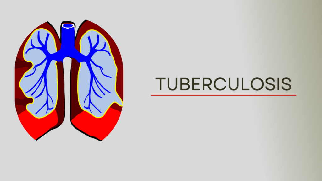
Tuberculosis
Tuberculosis
Definition : Tuberculosis is a chronic communicable, granulomatous disease caused by mycobacterium tuberculosis.
We have described in article :
Organisms responsible for tuberculosis
Management of pulmonary tuberculosis
Complications of pulmonary tuberculosis
Management of intestinal tuberculosis
Tubercular Meningitis

Organisms responsible for tuberculosis:
- Mycobacterium tuberculosis.
- Mycobacterium bovis.
- Mycobacterium africanum.
Primary pulmonary TB:
Primary TB refers to the infection of a previously uninfected (tuberculin-negative) individual.
Miliary Tuberculosis :
Blood-bome dissemination gives rise to miliary TB which may present acutely but more frequently is characterised by 2-3 weeks of fever, night sweats, anorexia, weight loss and a dry cough.
The classical appearances on chest X-ray are those of fine 1-2 mm lesions (millet seed’) distributed throughout the lung fields, although occasionally the appearances are coarser
Post primary pulmonary Tuberculosis:
Refers to the infection of a previously infected (tuberculin-positive) individual.
What is Ghon focus?
Ghon focus: The formation of a mass of granulomas surrounding an area of caseation leads to the appearance of the primary lesion in the lung, referred to as the Ghon focus.
What is Ghon complex?
Ghon Complex : The combination of a primary lesion and regional lymph node involvement is termed the Ghon Complex.
Management of pulmonary tuberculosis:
Clinical features:
Symptoms of pulmonary tuberculosis:
- Chronic cough, often with haemoptysis.
- Pyrexia of unknown origin
- Evening rise of temperature
- Night sweats
- Anorexia
- Weight loss, general debility
- Unresolved pneumonia
- May be asymptomatic (diagnosis on chest X-ray)
Signs of pulmonary tuberculosis:
- Raised temperature
- Patient may be cachectic.
- Cervical and other lymphadenopathy- When disseminated.
- Auscultation of chest is frequently normal.
- Features of pleural effusion may be found.
- Features of pneumothorax may be found.
- In advanced disease, widespread crackles may be found.
- Hepatosplenomegaly in disseminated TB.
Investigations of pulmonary tuberculosis:
- CBC, ESR & Hb %: High rise of ESR, lymphocytosis, anaemia
- Chest X-ray: Patchy opacity
- Sputum or bronchoalveolar lavage for AFB: May be found by Ziehl – Neelsen staining.
- PCR: Nucleic acid amplification
- Culture: (a)Solid media (Lowenstein-Jensen, Middlebrook). (b). Liquid media (eg BACTEC or MGIT).
- Tuberculin test: Useful only in primary or deep-seated infection.
- Biopsies of the pleura, lymph nodes and solid lesions within the lung (tuberculomas) may be required to confirm the diagnosis.
To know more about pulmonary tuberculosis click here
Treatment of Tuberculosis (According to WHO):
- New cases of smear-positive pulmonary tuberculosis, Severe extrapulmonary Tuberculosis, Severe smear-negative pulmonary TB, Severe concomitant HIV disease ==== Initial phase 2 months HRZE or 2 months HRZS, continuation phase 4 months HR
- Previously treated smear-positive pulmonary TB, Relapse, Treatment failure Treatment after default===== Initial phase 2 months HRZES+ 1 month HRZE, Continuation phase 5 months HRE.
H==== INH
R==== Rifampicin
E==== Ethambutol
Z==== Pyrazinamide
Pyridoxine (vitamin B6) is given at a dose of 10 mg daily to reduce the risk of isoniazid induced neuropathy
Causes of false positive MT test:
- Previous TB infection
- BCG vaccination
- Non-tubercular infection
- Old age
Causes of false negative MT (tuberculin) test:
- Severe TB (25% of cases negative)
- Newborn and elderly
- HIV (if CD4 count < 200 cells/ml)
- Recent infection (e.g. measles) or immunisation.
- Malnutrition
- Immunosuppressive drugs
- Malignancy
- Sarcoidosis
Complications of pulmonary Tuberculosis :
Pulmonary:
- Massive haemoptysis
- Fibrosis/emphysema
- Lung/pleural calcification
- Bronchiectasis
- Obstructive airways disease.
- Bronchopleural fistula
- Atypical mycobacterial infection.
- Cor pulmonale
- Aspergilloma
Non – Pulmonary :
- Empyema necessitans
- Laryngitis
- Enteritis
- Anorectal disease
- Amyloidosis
- Poncet’s polyarthritis
Indications of steroid (glucocorticoid) along with anti-TB drugs:
- Absolute indication: Bilateral adrenal tuberculosis (replacement therapy).
- Tubercular meningitis (to prevent adhesion).
- Tubercular pericarditis (to prevent adhesion).
- Tubercular pleural effusion (to prevent adhesion).
- Tubercular peritonitis (to prevent adhesion).
- Intestinal tuberculosis (to prevent adhesion and scarring).
Management of intestinal tuberculosis:
Clinical features of intestinal tuberculosis :
- Abdominal pain (acute or of several months duration).
- Low-grade fever.
- Night sweats
- Weight loss
- Features of anaemia (tiredness, palpitation).
- Alteration of bowel habit
- Features of intestinal obstruction-Constipation, vomiting
- Abdominal swelling-If ascites due to peritoneal involvement.
- Palpable abdominal mass, specially in right iliac fossa.
- Granulomatous hepatitis.
Investigations of intestinal tuberculosis:
- ESR-Raised.
- Barium follow through-Will show transverse ulceration, diffuse narrowing of the bowel with shortening of the caecal pole.
- USG and CT shows mesenteric thickening and lymph node enlargement.
- Serum alkaline phosphatase-Increased (suggests hepatic involvement).
- Confirmation is sought by endoscopy, laparoscopy or liver biopsy.
- Diagnosis is now possible on biopsy. specimens by rapid polymerase chain reaction techniques.
Treatment of intestinal tuberculosis :
- Chemotherapy with four drugs: Isoniazid, rifampicin, pyrazinamide & ethambutol.
- Treatment should last 1 year.
Management of Tubercular meningitis :
Clinical features:
Symptoms :
- Headache
- Vomiting
- Low-grade fever
- Lassitude
- Depression
- Confusion
- Behaviour changes
Signs:
- Meningism (Neck stiffness, Kernig’s sign)- Usually present, but may be absent.
- Oculomotor palsies
- Papilloedema
- Focal hemisphere signs
- Depression of conscious level
Investigations:
1) CSF examination:
- Pressure: Increased
- Appearance: It is usually clear but, when allowed to stand, a fine clot (‘spider web’) may form.
- Cell count and type: The fluid contains up to 5*10 cells/litre, predominamly lymphocytes.
- Protein: There is a rise in protein.
- Glucose: A marked fall in glucose
- Microbiology: Detection of the tubercle bacillus in a smear of the centrifuged deposit from the CSF may be difficult. The CSF should be cultured but as this result will not be known to 6 weeks, treatment must be started without waiting for confirmation.
2) Brain imaging: May show hydrocephalus, brisk meningeal enhancement on enhanced CT and/or an intracranial tuberculoma.
Treatment of tubercular meningitis :
- Anti-TB drugs: Initial 2 months, 4 drugs regimen (isoniazid, rifampicin, ethambutol & pyrazinamide) Continuation by 2 drugs (isoniazid & rifampicin). Treatment should be continued for 1 year.
- Corticosteroid-Improves mortality but not focal neurological damage.
- Surgical ventricular drainage may be needed if obstructive hydrocephalus develops.
Stages of tubercular meningitis:
- Stage 1 (early): Non-specific symptoms and signs without alteration of consciousness.
- Stage 11 (Intermediate): Altered consciousness without coma or delirium + minor focal neurological signs.
- Stage III (advanced): Stupor or coma, severe neurological deficits, seizure or abnormal movements.

Your point of view caught my eye and was very interesting. Thanks. I have a question for you.